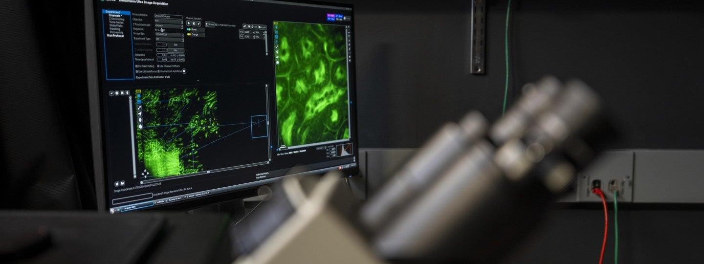
Microscopy & Image Analysis Workstations

About the GC3F Imaging Facility

The light microscopy center within the GC3F located in 270 Klamath Hall is an interdisciplinary facility which seeks to provide centralized space, personnel, and technical tools toward advancing optical imaging in the biological sciences. The facility is designed to serve as a conduit for state of the art microscopy and image analysis resources to the University of Oregon, Oregon State University, and Oregon Health and Science University communities. We offer hands-on microscopy training, experimental guidance, and image analysis training and support.

Acknowledgements and Support

Continual support for core facilities relies in part on proper acknowledgements in publications.
If the services offered by the imaging facility have been instrumental in your research, please acknowledge the GC3F in your publications and let us know!

____________________________________________________________________________________________

The Core currently offers training and support for the following equipment:

Leica SPE Laser Scanning Confocal and Widefield

Cytiva DeltaVision Ultra - Widefield with Deconvolution

Zeiss LSM 880 with Elyra Structured Illumination and Airy Scanning

Nikon CSU-W1 SoRa Spinning Disk

Amersham Typhoon scanner

Zeiss LSM 710 with Elyra Structured Illumination and Airy Scanning (Knight Campus)

Cherry Temp System

Tokai Hit Environmental Chamber

Analysis Workstations

Equipment currently in development:

Custom-built Light Sheet microscope


Leica SPE Laser Scanning Confocal and Widefield

Back to Top

  • 405nm, 488nm, 561nm, 633nm laser excitation lines, and DIC illumination
  • DAPI, FITC, TRITC equivalent for widefield imaging
  • Motorized stage, upright body
  • 5x, 10x, 25x water dipping, 40x oil, 40x water dipping, 63x oil objectives
  • Acquisition software is LAS X on Windows 10
  • Usage Rates - Internal (UO, OSU, OHSU): $20/hr, External $28/hr


Cytiva DeltaVision Ultra - Widefield with Deconvolution

Back to Top

  • LED excitation lines of 405nm, 458nm, 488nm, 514nm, 555nm, 594nm, 633nm
  • 20x LWD, 20x, 60x oil, 100x oil objectives
  • Motorized stage, pco.edge sCMOS camera, inverted body
  • Acquisition software is Acquire Ultra on Linux CentOS 7
  • Usage Rates - Internal (UO, OSU, OHSU): $20/hr, External: $28/hr


Zeiss LSM 880 with Elyra Structured Illumination and Airy Scanning

Back to Top

  • 405nm, 458nm, 488nm, 514nm, 561nm, 633nm laser excitation lines
  • 10x, 20x, 40x water immersion, 40x oil, 63x oil objectives
  • Motorized stage, light-tight enclosure, inverted body
  • Acquisition Software is Zen Black on Windows 7
  • Usage Rates - Internal (UO, OSU, OHSU): $33/hr, External: $48/hr


Nikon CSU-W1 SoRa Spinning Disk

Back to Top

  • Spinning Disk excitation lines are 405nm, 488nm, 561nm, 633nm, and DIC illumination
  • TIRF imaging with excitation lines of 488nm, 561nm, and 633nm
  • Digitial Multi-Mirror Device and Lumencor Spectra X for photo-stimulation with LED stimulation lines of 390nm, 440nm, 488nm, 561nm, 594nm, 633nm, 740nm
  • 405nm photobleaching laser line
  • 4x, 10x, 20x, 40x water immersion, 60x water immersion, 60x oil TIRF objectives
  • Motorized stage, dual Prime BSI sCMOS cameras for optional simultaneous green/red imaging, inverted body
  • Acquisition sofware is Nikon Elements + JOBs on Windows 10
  • Usage Rates - Internal (UO, OSU, OHSU): $36/hr, External $53/hr


Amersham Typhoon scanner

Back to Top

  • fluorescent imaging with multiple lasers (Cy2, Cy3, Cy5)
  • imaging of storage phosphor screens
  • densitometric imaging
  • Usage Rates - Internal (UO, OSU, OHSU): free to use


Zeiss LSM 710 with Elyra Structured Illumination and Airy Scanning (Knight Campus)

Back to Top

  • 405nm, 458nm, 488nm, 514nm, 561nm, 633nm laser excitation lines
  • 20x LWD, 63x oil, 100x oil objectives
  • Motorized stage, light-tight enclosure, inverted body
  • Acquisition Software is Zen Black on Windows 7
  • Usage Rates - Internal (UO, OSU, OHSU): $33/hr, External: $48/hr


Cherry Temp System

Back to Top

  • enables temperature shifts from 5-45 celsius degrees in 10 seconds with a 0.1 degree error.
  • Usage Rates - Internal (UO, OSU, OHSU): free to use


Tokai Hit Environmental Chamber

Back to Top

  • portable stage top incubation system
  • Usage Rates - Internal (UO, OSU, OHSU): free to use


Support and training will exist in the near future for:

Back to Top

  • Custom-built Light Sheet microscope

          If you have a project that you think may benefit from light sheet imaging, please contact Adam Fries at afries2@uoregon.edu.


The Core currently has 4 analysis workstations and include the following software:

Back to Top

(Usage Rates - Internal (UO, OSU, OHSU): $25/hr, External $37/hr)

  • Imaris 9.9 with Cell and Filaments Modules
  • Imaris 9.9 Converter and Stitcher
  • Prism8
  • Matlab 2018a
  • Adobe Illustrator and Photoshop
  • Microsoft Office
  • Zen Blue/Black
  • Python3
  • FIJI

Calendar Access and Training:

Back to Top

  • Instrument reservations are made though iLab and each user will need to register an iLab account if they do not have one. Once training on the instrument has been scheduled and completed, new users will be granted calendar access.


  • Individualized training recharge for each of the instruments is a onetime fee of $150 ($300 for external customers).


  • Contact Adam Fries afries2@uoregon.edu for more information regarding instruments, training, or analysis. Or visit the office in Klamath 270D.


----------------------------------------------------------------------------------------------------------------


(If you have urgent microscopy-related questions in the meantime, please email afries2@uoregon.edu or dturnbul@uoregon.edu)