The light microscopy center within the GC3F offers training and support for the following equipment:
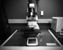
Leica SPE Laser Scanning
Confocal and Widefield
- 405 nm, 488 nm, 561 nm, 633 nm laser excitation lines, and DIC illumination
- DAPI, FITC, TRITC equivalent for widefield imaging
- Motorized stage, upright body
- 5x, 10x, 25x water dipping, 40x oil, 40x water dipping, 63x oil objectives
- Acquisition software is LAS X on Windows 10
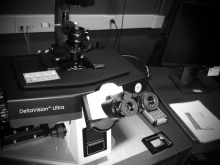
Cytiva DeltaVision Ultra
Widefield with Deconvolution
- LED excitation lines of 405 nm, 458 nm, 488 nm, 514 nm, 555 nm, 594 nm, 633 nm
- 20x LWD, 20x, 60x oil, 100x oil objectives
- Motorized stage, pco.edge sCMOS camera, inverted body
- Acquisition software is Acquire Ultra on Linux CentOS 7
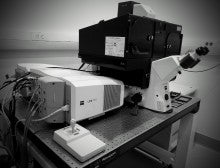
Zeiss LSM 880
With Elyra Structured Illumination and AiryScan I
- 405 nm, 458 nm, 488 nm, 514 nm, 561 nm, 633 nm laser excitation lines
- 10x, 20x, 40x water immersion, 40x oil, 63x oil objectives
- Motorized stage, light-tight enclosure, inverted body
- Acquisition Software is Zen Black on Windows 7

Nikon CSU-W1 SoRa Spinning Disk
- Spinning Disk excitation lines are 405 nm, 488 nm, 561 nm, 633 nm, and DIC illumination
- TIRF imaging with excitation lines of 488 nm, 561 nm, and 633 nm
- Digitial Multi-Mirror Device and Lumencor Spectra X for photo-stimulation with LED stimulation lines of 390 nm, 440 nm, 488 nm, 561 nm, 594 nm, 633 nm, 740 nm
- 405 nm photobleaching laser line
- 4x, 10x, 20x, 40x water immersion, 60x water immersion, 60x oil TIRF objectives
- Motorized stage, dual Prime BSI sCMOS cameras for optional simultaneous green/red imaging, inverted body
- Acquisition software is Nikon Elements + JOBs on Windows 10
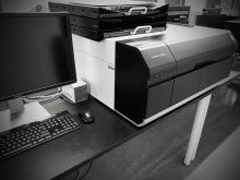
Amersham Typhoon Scanner
- Fluorescent imaging with multiple lasers (Cy2, Cy3, Cy5)
- Imaging of storage phosphor screens
- Densitometric imaging
Zeiss LSM 710 (down for repair)
With Elyra Structured Illumination and Airy Scanning
This is located in the Knight Campus.
- 405 nm, 458 nm, 488 nm, 514 nm, 561 nm, 633 nm laser excitation lines
- 20x LWD, 63x oil, 100x oil objectives
- Motorized stage, light-tight enclosure, inverted body
- Acquisition Software is Zen Black on Windows 7
Cherry Temp System
- Enables temperature shifts from 5–45 Celsius degrees in 10 seconds with a 0.1 degree error.

Tokai Hit Environmental Chamber
- Portable stage top incubation system
Custom-built Light Sheet Microscope
If you have a project that you think may benefit from light sheet imaging, please contact Adam Fries at afries2@uoregon.edu.
Workstations
GC3F has four analysis workstations available to reserve.
Contact Adam Fries for more information regarding instruments, training, or analysis, or visit the office in Klamath 270D.